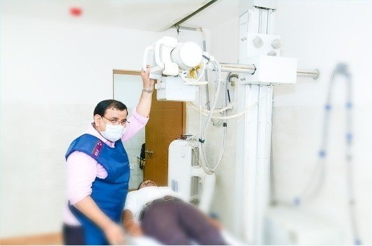
DIGITAL X-RAY
An x-ray is a noninvasive medical test that helps physicians diagnose and treat medical conditions. Imaging with x-ray involves exposing a part of the body to a small dose of radiation for producing pictures of the inside of a body. X-rays is the oldest and most frequently used form of medical imaging. X-rays give a lot of information about the bones within the body and the advancement in x-ray technology over the years have made it a good diagnostic tool even for soft tissues information.
Tests done on Digital X-Ray
At Narayan Swaroop Hospital, we perform a variety of x-rays for various parts of the body and organs. A list of such scans are as follows: Chest X-ray, X-ray Spine, X-ray Knee Joint, X-ray Hip Joint, X-ray small joints, X-ray KUB, Intravenous pyelography (IVP), Baruim Studies, Fistulogram, Sinogram, Hysterosalpingography (HSG).
At Narayan Swaroop Hospital we have one of the most advanced digital x-ray imaging and the report is handed over to the patient instantly, making it easy for the patient to start his/her treatment based on the diagnosis.
Digital Radiography
Digital radiography is one of the recent advances in x-ray imaging, where digital x-ray sensors are used instead of traditional photographic film. Advantages of digital imaging include time efficiency, ability to digitally transfer and enhance images and reduced radiation. Instead of x-ray film, digital radiography uses a digital image capture device. This gives us the advantage of immediate image preview and elimination of costly film processing steps. A wider dynamic range, which makes it more forgiving for over-exposure and under-exposure as well as the ability to apply special image processing techniques that enhances overall display of the image. X-ray is one of the basic and important imaging tools.
What is an X-Ray
X-ray is the most common imaging test used for detecting and diagnosing medical conditions. X-rays have been in use by the doctors for many decades. X-ray helps in looking at the inside of the body without having to cut open the body or make an incision. In this way an x-ray helps in diagnosing, monitoring, and treating many medical conditions. X-ray carries very minor risks and the benefits of the x-rays outweigh its risks. There are different types of x-rays used for different purposes. For example, barium enema is an x-ray which is done to look at your gastrointestinal tract and mammogram is a type of x-ray done to examine the breasts.
HSG – Hysterosalpingography
Hysterosalpingography is an x-ray examination of a woman’s uterus and fallopian tubes that uses a special form of x-ray called fluoroscopy and a contrast material.
An x-ray (radiograph) is a noninvasive medical test that helps physicians diagnose and treat medical conditions. Imaging with x-rays involves exposing a part of the body to a small dose of ionizing radiation to produce pictures of the inside of the body.
X-rays are the oldest and most frequently used form of medical imaging. Fluoroscopy is a special x-ray technique that makes it possible to see internal organs in motion. During a hysterosalpingogram, the uterus and fallopian tubes are filled with a water-soluble contrast material and the radiologist is able to use fluoroscopy to view and assess their anatomy and function.
This procedure should not be performed if you have an active inflammatory condition. Tell your physician or technologist if you have a chronic pelvic infection or an untreated sexually transmitted disease. Wear loose, comfortable clothing and leave jewelry at home. You may be asked to wear a gown.
Procedure
The procedure can be used to investigate repeated miscarriages that result from congenital or acquired abnormalities of the uterus.
Hysterosalpingography is used to evaluate the openness of the fallopian tubes and to monitor the effects of tubal surgery, including:
Blockage of the fallopian tubes due to infection or scarring, Tubal ligation.
The closure of the fallopian tubes in a sterilization procedure and a sterilization reversal.
The closure of the fallopian tubes in a sterilization procedure and a sterilization reversal. The re-opening of the fallopian tubes following a sterilization or disease-related blockage.
How does the procedure work
X-rays are a form of radiation like light or radio waves. X-rays pass through most objects, including the body. Once it is carefully aimed at the part of the body that is to be examined, an x-ray machine produces a small burst of radiation that passes through the body, recording an image on photographic film or a special detector.
Fluoroscopy uses a continuous or pulsed x-ray beam to create a sequence of images that are projected onto a fluorescent screen, or television-like monitor. When used with a contrast material, which clearly defines the area being examined by making it appear dark (or by electronically reversing the image contrast to white), this special x-ray technique makes it possible for the physician to view joints or internal organs in motion. Still images or movies are also captured and stored electronically on a computer.
Until recently, x-ray images were maintained on large film sheets (much like a large photographic negative). Today, most images are digital files that are stored electronically. These stored images are easily accessible for diagnosis and disease management.
Experience during and after the procedure
This examination causes only minor discomfort. Sedation or anaesthesia might be used to minimize the pain experienced during the examination.
There may be slight discomfort and cramping when the catheter is placed and the contrast material is injected, but it does not last long.
Benefits vs. Risks
Benefits
- Hysterosalpingography is a minimally invasive procedure with rare complications.
- Hysterosalpingography is a relatively short procedure that can provide valuable information on a variety of abnormalities that cause infertility or problems in carrying a fetus to term.
- Hysterosalpingography can occasionally open fallopian tubes that are blocked allowing the patient to become pregnant afterwards.
- No radiation remains in a patient’s body after an x-ray examination.
- X-rays usually have no side effects in the typical diagnostic range for this exam.
- Hysterosalpingography is primarily used to examine women who are facing difficulty in getting pregnant by allowing the radiologist to evaluate the shape and structure of the uterus, the openness of the fallopian tubes and any scarring within the uterine or peritoneal (abdominal) cavity.
RISK
- There is always a slight chance of cancer from excessive exposure to radiation. However, the benefit of an accurate diagnosis far outweighs the risk.
- In the event of a chronic inflammatory condition, pelvic infection or untreated sexually transmitted disease, be certain to notify the physician or technologist before the procedure to avoid worsening of infection.
- Women should always inform their physician or x-ray technologist if there is any possibility that they are pregnant.